 YouTube Channel
Microlaryngoscopy with excisional biopsy involves surgically removing a small piece of tissue from the vocal cords or larynx to be examined under a microscope. It is used when an unknown lesion or abnormality, such as a nodule, mass, or ulcer, is present and a precise diagnosis is needed.
This procedure is done under general anesthesia using specialized tools through the mouth. Through a microscope, the surgeon views the vocal cords and carefully removes the target tissue. Afterward, patients are typically advised to rest their voice and may feel mild throat discomfort. Results from the biopsy usually guide further treatment. The risk of complications is low, and speech is usually unaffected once healing is complete, though some patients may need follow-up procedures or therapy based on the biopsy results.
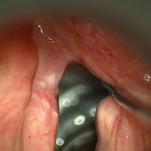
Before: Microlaryngoscopy removal of Leukoplakia
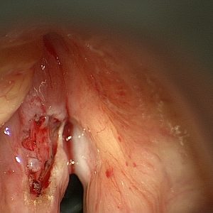
After: Microlaryngoscopy removal of Leukoplakia